Product Description
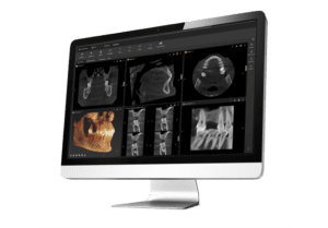
DTX Studio™ suite connects the devices and technologies in your dental practice or lab – in one single platform.
QUICKcompose™ for fast image review, appearing automatically following the scan
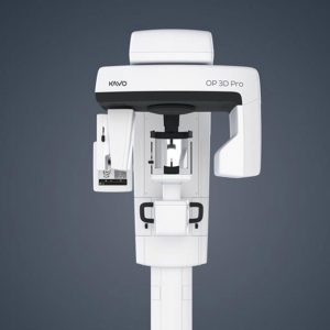
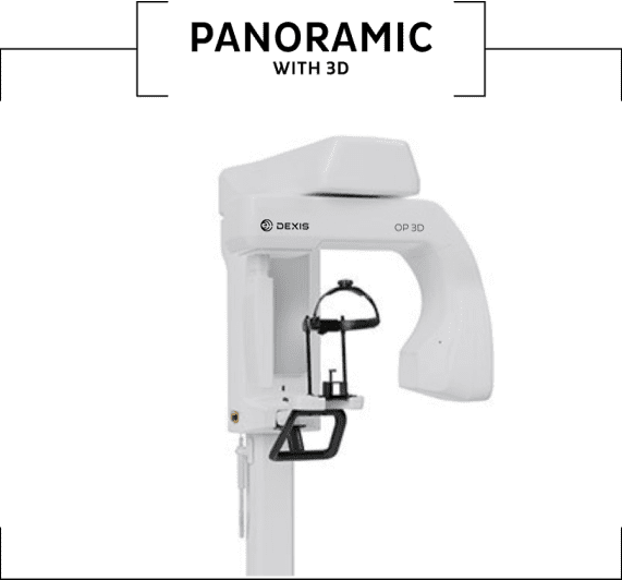
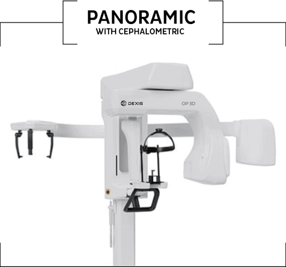
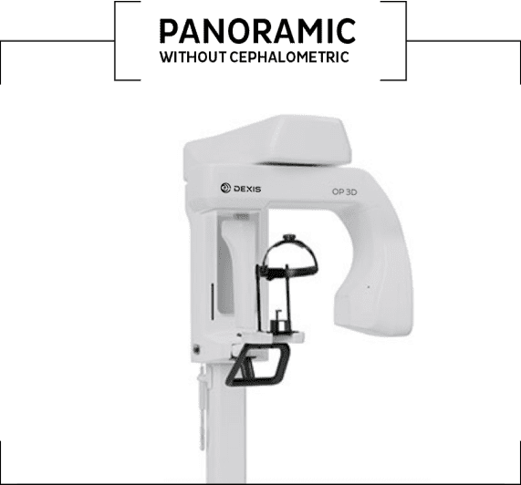
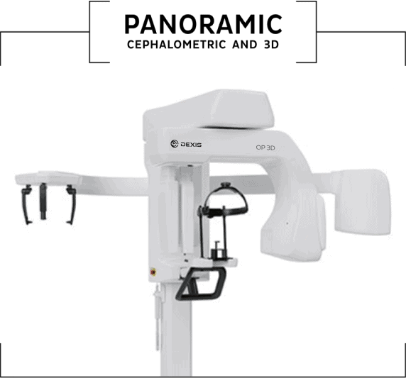
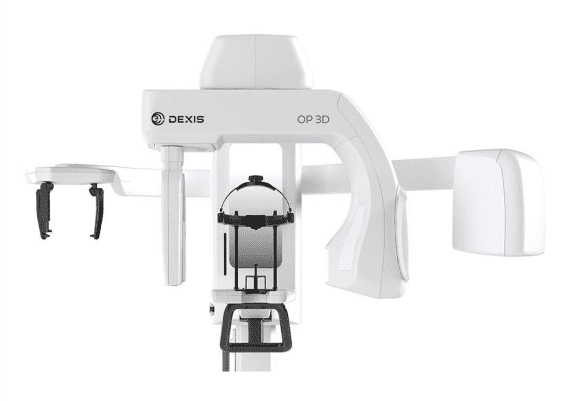
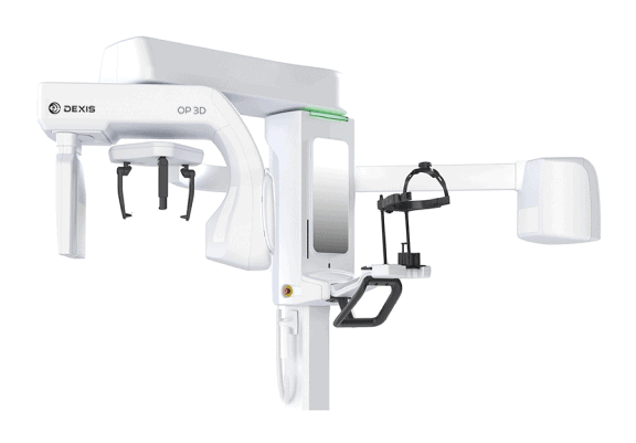
Configurable device platform: Panoramic, Cephalometric and 3D imaging
Optimized imaging workflows and lead-free device
Designed for efficiency
Every feature of the DEXIS OP 3D is designed to increase practice efficiency. Preparing the device for a scan is fast with an easy patient positioning system and intuitive graphical user interface. All imaging protocols are optimized for practice workflows.
Intuitive operation, connected to the future
All functions can be easily and intuitively controlled in a time-saving way via your laptop or PC through the practice’s local network. Only the patient positioning is set on the device.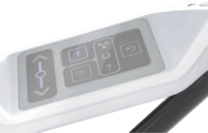
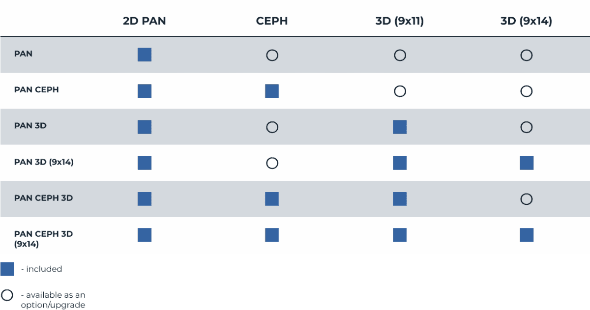
Configuration
3D
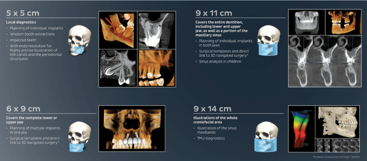
- 4 resolutions for 3D (Low Dose Technology™ (LDT), Standard, High, Endo) combined with Metal Artifact Reduction (MAR) technology
- 4 predefined volumes: 5×5, 6×9, 9×11 and (optional) 9×14 cm – thanks to SMARTVIEW™ 2.0 the volumes are freely positionable and height adjustable in 5 mm steps between 5 and 9 cm before the exposure, leading up to 36 possible FOV sizes in total.
Panoramic
- Fast Scan – 2D panoramic imaging in just 9 seconds
- ORTHOfocus™ feature for providing the optimum panoramic image layer automatically
- Panoramic programs for covering the daily needs of a busy practice
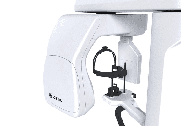
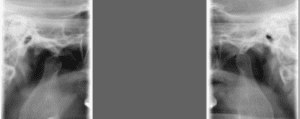
Cephalometric
- Innovative and patented ORTHOceph™ Plus design with fast cephalometric imaging scan times and adjustable field sizes for perfect image quality with minimal dose
The DEXIS OP 3D can grow with your clinical needs
The DEXIS OP 3D is designed to be upgradeable, allowing it to grow with the needs of your practice. The cephalometric or 3D imaging capabilities can be added also later on.
Features
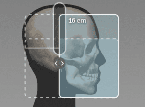
Four predefined 3D volume diameters plus the possibility to customize the volume size
The four predefined FOVs of the DEXIS OP 3D are based on true clinical needs and adjustable in height. FOV 5×5 with its endo resolution is optimised for single-tooth and localized diagnostics. FOV 6×9 offers the capability of scanning either the lower or upper jaw, whereas FOV 9×11 combines both. With the largest FOV 9×14, TMJs can be conducted.
Metal Artifact Reduction (MAR): To provide optimum image quality, the Metal Artifact Reduction (MAR) is activated with all FOV sizes and resolutions of the DEXIS OP 3D. MAR is optimized to assist in all cases ranging from endodontics and implants planning to maxillofacial imaging.
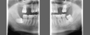
PAN
Panoramic images with automatically selected optimum layer – ORTHOfocus
Programs to fit your clinical needs: Standard, pediatric and segmented panoramics along with bitewing and lateral-TMJ programs are included to cover the panoramic imaging needs of a busy practice. With the ORTHOfocus feature, the optimum panoramic image layer is automatically obtained, enabling forgiving patient positioning. The result is consistent image quality every time.
The pediatric panoramic program has a clinically adapted image layer and reduced image height.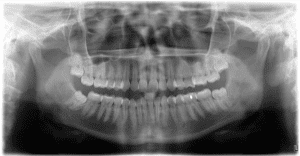
The bitewing program provides a quick and easy alternative to intraoral bitewing imaging.
The TMJ program provides a lateral view of temporomandibular joints, with an open or closed mouth.
QUICKcompose feature: fast image review
Available for panoramic, cephalometric and 3D modalities, the QUICKcompose feature offers a quick preview of the captured image, allowing a timely evaluation. The image appears on the graphical user interface automatically as soon as the scan is completed.
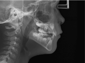
Cephalometric imaging for all your clinical needs
The innovative, patented ORTHOceph Plus design of the DEXIS OP 3D takes cephalometric imaging workflow to a new level. The DEXIS OP 3D provides all needed protocols such as lateral and pediatric lateral projections with adjustable field widths, posterior-anterior (PA) projections and carpus (carpus holder is optional) imaging — with fast scan times and a minimal dose. All combined with an intuitive graphical user interface and automated sensor movements to enable smooth workflows.
Lateral cephalometric images provide rich anatomical details with exceptional visibility of the soft tissue borderline.